Home
/ Neuron Diagram Labeled, A Labelled Diagram Of Neuron With Detailed Explanations, The below mentioned article provides a study note on the neurons as structural and functional unit of neural system.
Neuron Diagram Labeled, A Labelled Diagram Of Neuron With Detailed Explanations, The below mentioned article provides a study note on the neurons as structural and functional unit of neural system.
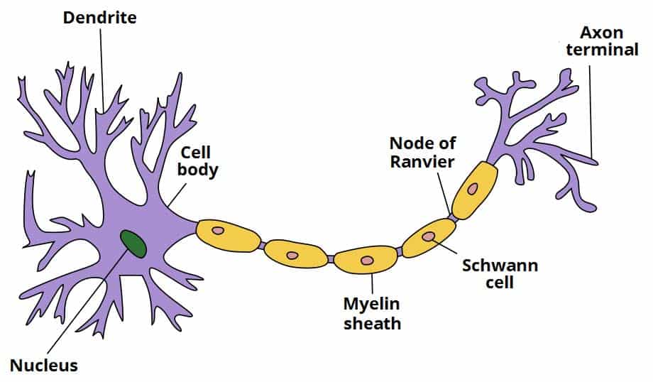
Neuron Diagram Labeled, A Labelled Diagram Of Neuron With Detailed Explanations, The below mentioned article provides a study note on the neurons as structural and functional unit of neural system.. Choose the correct names for the parts of the neuron. A neuron is a structural and functional unit of the neural tissue and hence the neural system. Axon, cell body, dendrites, nucleus, terminal. This neuron part contains instructions for making. Please send your queries to ncerthelp@gmail.com you can aslo visit our facebook page to get quick help.
Image 48129376 discover millions of stock images, photos, video and audio. Label the parts of the neuron in the diagram. Parts of a neuron diagram although they have a characteristic elongated shape, they vary widely in size and properties based on their location and type of functions they perform. The principles outlined above can be applied to the neuron and its ionic contents. Draw a labelled diagram of a neuron.
Ultrastructure Of Nerves Classification Neurones Teachmeanatomy from teachmeanatomy.info Plasticity the brain's ability to recover from brain/nerve damage by possibly creating new pathways for previous messages action potential this allows messages to flow from neuron to neuron as an electrical charge is created when positively charged sodium ions flow into a neuron and flows out as positively charged potassium charges. Based on it, they need to draw a. A group of neurons forms a nerve. Axon terminals the hair like ends of the axon cell body the cell body of the neuron. These are called dendrites, and they carry incoming messages to the cell. Diagram of neuron anatomy free vector. Labeled neuron diagram neurons are the basic organizational units of the brain and nervous system. Formed by the schwann cells in pns, 20% protein, and 80% lipid.
Formed by the schwann cells in pns, 20% protein, and 80% lipid.
Unlabeled diagram of nervous system. Plasticity the brain's ability to recover from brain/nerve damage by possibly creating new pathways for previous messages action potential this allows messages to flow from neuron to neuron as an electrical charge is created when positively charged sodium ions flow into a neuron and flows out as positively charged potassium charges. Insulating layer around a nerve fiber. Read the definitions, then label the neuron diagram below. An axon is a long, thin fiber that extends from the nerve cell body. Alex bolano on may 29,. When a nerve impulse reaches this knob, a drug called a neurotransmitter is released from vesicles into the synapse the neurotransmitter diffuses across the gap and binds to receptors on the membrane of the adjacent neuron or muscle cell. 3.1 how to draw a neuron diagram from sketch step 1: Neurons pass messages to each other using a special type of electrical signal. The students may follow these steps to make their neuron diagram, but the process is complex: Label the parts of the neuron in the diagram. The nucleus of the neuron is found in the soma. Labeled diagram of the neuron, nerve cell that is the main part of the nervous system.
An axon is a long, thin fiber that extends from the nerve cell body. While they have the common features of a typical cell, they are structurally and functionally unique from other cells in many ways. Dendrites receive messages from other neurons. Anatomy and physiology questions and answers. Diagram of neuron with labels here is the description of human neuron along with the diagram of the neuron and their parts.
Neuron Basic Structure And Functions from www.getbodysmart.com Diagram of neuron anatomy free vector. This neuron part gives messages to muscle tissue. Many spiderlike arms branch from the cell body. An axon is a long, thin fiber that extends from the nerve cell body. Label the parts of the neuron in the diagram. Human anatomy diagrams show internal organs, cells, systems, conditions, symptoms and sickness information and/or tips for healthy living. Dendrites receive messages from other neurons. These are called dendrites, and they carry incoming messages to the cell.
Shutterstock customers love this asset!
From there the message can move to the next neuron. Various processes (appendages or protrusions) extend from the cell body. Not all terms will be used. This labelled diagram quiz on neuron is designed to assess your basic knowledge in structure and function of neuron. Axon terminals the hair like ends of the axon cell body the cell body of the neuron. Certain neurons may almost equal the length of body. An axon is a long, thin fiber that extends from the nerve cell body. A neuron communicates with other neurons at special places called synapses or synaptic clefts. This vast connectional architecture explains that computational complexity of the human brain. First, the students need to draw a circle. 3.1 how to draw a neuron diagram from sketch step 1: Read the definitions, then label the neuron diagram below. There is a printable worksheet available for download here so you can take the quiz with pen and paper.
Where is the cell membrane on a neuron. Neurons communicate with each other by generating and. Read the definitions, then label the neuron diagram below. This diagram depicts neuron anatomy.human anatomy diagrams show internal organs, cells, systems, conditions, symptoms and sickness information and/or tips for healthy living. The principles outlined above can be applied to the neuron and its ionic contents.
Labeled Diagram Of The Neuron from thumbs.dreamstime.com From there the message can move to the next neuron. Read the definitions, then label the neuron diagram below. The principles outlined above can be applied to the neuron and its ionic contents. The message then moves through the axon to the other end of the neuron, then to the tips of the axon and then into the space between neurons. The neurons are of three kinds according to their functions. This is an online quiz called label a neuron. Image 48129376 discover millions of stock images, photos, video and audio. Axon terminals the hair like ends of the axon cell body the cell body of the neuron.
Anatomy and physiology questions and answers.
An axon is a long, thin fiber that extends from the nerve cell body. The principles outlined above can be applied to the neuron and its ionic contents. Labeled neuron diagram neurons are the basic organizational units of the brain and nervous system. This is an online quiz called label a neuron. Shutterstock customers love this asset! Not all terms will be used. Where is the cell membrane on a neuron. Unlabeled diagram of nervous system. Various processes (appendages or protrusions) extend from the cell body. Node of ranvier soma answer bank axon terminal glial cell axon nerve ganglion dendrite nucleus. Read the definitions, then label the neuron diagram below. Choose the correct names for the parts of the neuron. Parts of a neuron diagram although they have a characteristic elongated shape, they vary widely in size and properties based on their location and type of functions they perform.